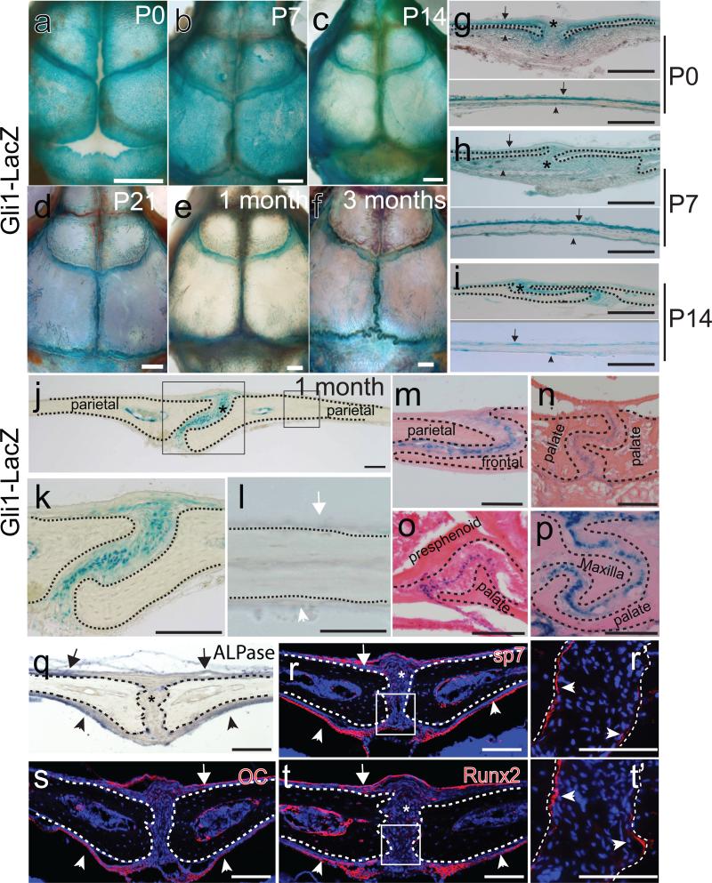
Figure 1.
Gli1+ cells are restricted to the suture mesenchyme of craniofacial bones and are undifferentiated cells. (a-f) Whole mount LacZ staining (blue) of calvarial bones of newborn (P0), 7, 14 and 21 day old (P7, P14, P21) and one- and three-month-old Gli1-LacZ mice. (g-i) LacZ staining of sections of sagittal sutures and parietal bones of P0, P7 and P14 mice indicates Gli1+ cells are present in the suture mesenchyme (asterisks), periosteum (arrows) and dura (arrowheads). (j-l) LacZ staining of sections of the sagittal suture of one-month-old Gli1-LacZ mice. Asterisk indicates exclusive Gli1 expression within the suture mesenchyme. No positive staining is detectable in the periosteum (white arrow) and dura (white arrowhead). Boxed areas in j are displayed in k and l. (m-p) LacZ staining of the mid-suture mesenchyme in the coronal (m), interpalatal (n), presphenoid-palatal (o) and maxilla-palatal (p) sutures of one-month-old Gli1-LacZ mice. (q-t) ALPase (blue) and immunohistochemical (red) staining of sagittal sutures of one-month-old mice. Osteogenic markers including ALPase (q), Sp7 (r), osteocalcin(OC) (s) and Runx2 (t) are not detectable in the suture mesenchyme (asterisks). The periosteum (arrows) and dura (arrowheads) strongly express these markers. Boxed areas in r and t are enlarged in panels r’ and t’, respectively, showing positive expression in the osteogenic fronts (arrowheads). Dotted lines outline margins of craniofacial bones. Scale bars in panels a-f, 1mm; other scale bars, 100 μm.

