Figure 2.
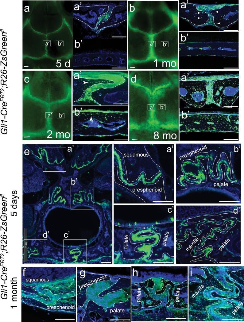
Gli1+ cells in the suture mesenchyme contribute to adult craniofacial bone turnover. Gli1-CE;R26ZsGreenfl mice were induced at 1 month of age with tamoxifen. (a-d) Gli1+ cells in the adult sagittal suture mesenchyme 5 days and 1, 2 and 8 months after induction. Arrowheads indicate positively labeled osteocytes. Boxes indicate the approximate positions of sections shown in the right panels (a' and b'). (e-i) Five days (e, a'-d') and one month (f-i) after induction, fluorescently labeled cells are detectable in the craniofacial sutures (low magnification image in e) including the squamous-presphenoid (panels a', f), presphenoid-palatal (panels b', g), interpalatal (panels c', h) and maxilla-palatal (panels d', i) sutures. Dotted lines outline craniofacial bones. Scale bars in panel a-d, 1mm; other scale bars, 100 μm.

