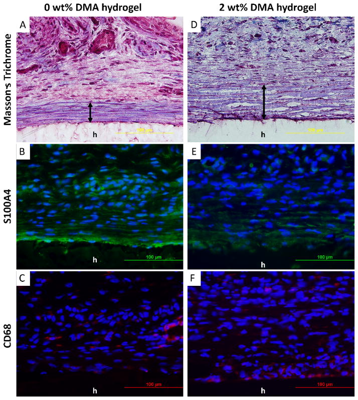
Figure 8.
Masson’s trichrome (A, D) and immunofluorescent (B, C, E, F) staining of 0 and 2 wt% DMA hydrogel and surrounding tissue after 4 week of subcutaneous implantation. Cell nuclei, fibroblasts, and macrophages were stained by DAPI (blue), S100A4 (green), and CD68 (red), respectively. h : hydrogel; double-headed arrows : fibrous capsule.

