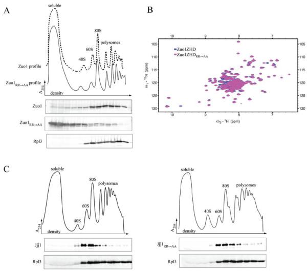
Fig. 2.
Zuo1R247,251A (Zuo1RR→AA) is largely dissociated from ribosomes. A) Lysate from Δzuo1 cells expressing wild-type Zuo1 or Zuo1RR→AA from the native promoter was centrifuged through a sucrose gradient to separate ribosomal subunits, monosomes and polysomes. Fractions were collected. Upper, absorbance at 254 nm plotted versus the relative time of fraction collection (density); dotted line (Zuo1), solid line (Zuo1RR→AA). Lower, fractions were analyzed by immunoblotting using antibodies specific for Zuo1 and Rpl3 (Rpl3 distribution in both experiments was indistinguishable, the WT sample is shown for reference). B) 2D 15N-1H Heteronuclear Single-Quantum Correlation (HSQC) NMR spectra of WT Zuo1166-303 (Zuo1ZHD), which includes the Zuotin homology domain (ZHD), and Zuo1ZHDRR→AA. C) Lysate from Δjjj1 cells expressing wild-type Jjj1 (left) or Jjj1RR→AA (right) from the native promoter was subjected to sucrose gradient analysis as in (A). Upper, absorbance at 254 nm plotted versus the relative time of fraction collection (density). Lower, fractions were analyzed by immunoblotting using antibodies specific for the C-terminus of Jjj1 and Rpl3.

