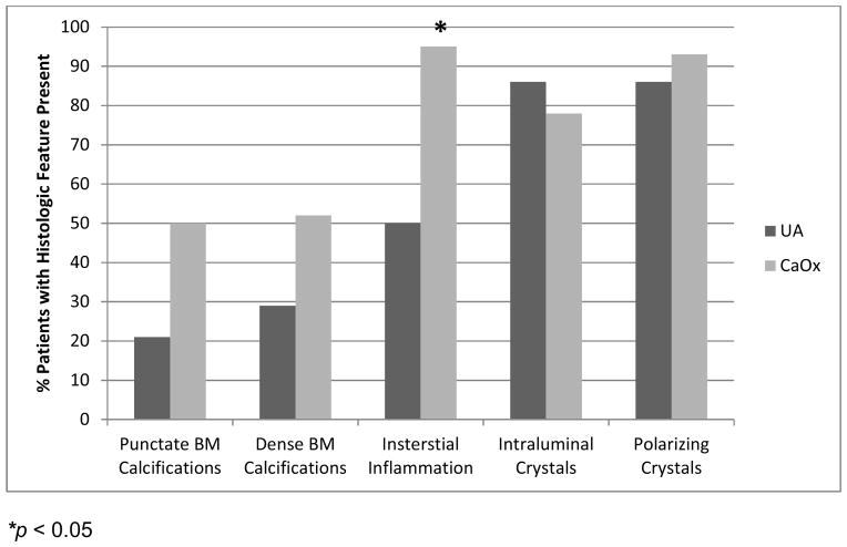
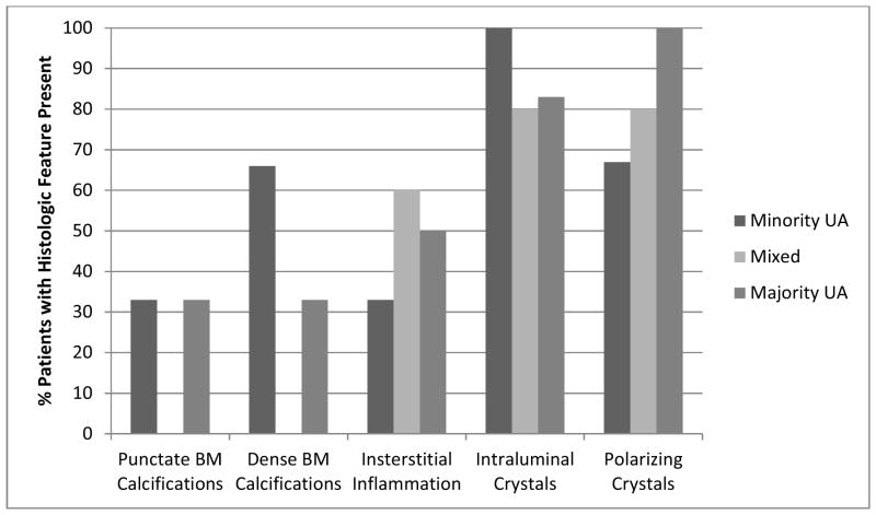
Figure 3.
Quantification of plaque and plug in papillary biopsies. Panel A: CaOx patients had more interstitial inflammation than UA patients and tended towards more punctate calcifications. Panel B: UA stone formers with majority CaOx (minority UA) tended to have more dense calcifications than the others, although this did not achieve statistical significance


