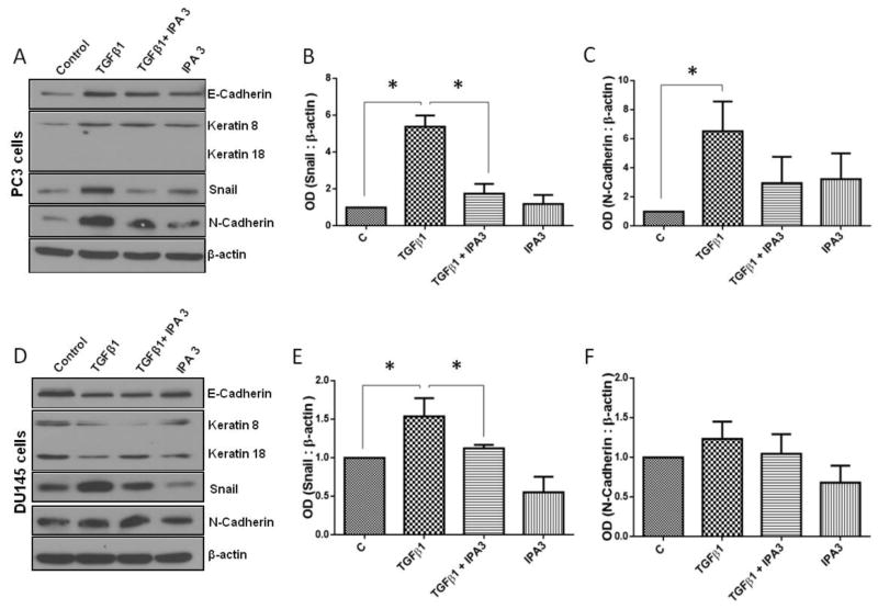
Figure 6. IPA 3, Selective Pak1 inhibitor inhibits TGFβ1-mediated EMT in prostate cancer cells.
A, Representative Western blot images of EMT marker expression in PC3 cells following 72 h treatments with TGFβ1 (5 ng/ml), IPA 3 (15 μM), or combination, with treatments repeated every 24 hours. B and C. Bar graph representing optical densitometry for mesenchymal markers, Snail and N-Cadherin expressions respectively in PC3 cells following 72 h treatments with TGFβ1 (5 ng/ml), IPA 3 (15 μM), or combination (n= 3). D, Representative Western blot images of EMT markers expression in DU145 cells following 72 h treatments with TGFβ1 (5 ng/ml), IPA 3 (15 μM), or combination. E and F. Bar graph representing optical densitometry of mesenchymal markers, Snail and N-Cadherin expressions respectively in DU145 cells following 72 h treatments with TGFβ1 (5 ng/ml), IPA 3 (15 μM), or combination (n= 3). *p<0.05, Data presented as Mean ± SEM.

