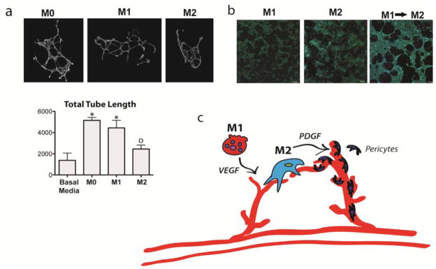
Figure 3. Contributions of M1 and M2 macrophages to angiogenesis.

(a) M1 but not M2 macrophages increase sprouting of endothelial cells on Matrigel (* indicates p<0.05 and o indicates p>0.05 compared to control, one-way ANOVA and Tukey’s post-hoc analysis). Networks were stained with Live/Dead kit (Invitrogen), imaged at 10x magnification with an Olympus IX81 fluorescent microscope, and analyzed with ImageJ’s Angiogenesis Analyzer macro. (b) Endothelial cells organized into a loosely connected network on fibrin gel when the media is changed from M1-conditioned media to M2-conditioned media, which did not occur when endothelial cells were cultured uniformly in either M1- or M2-conditioned media. Endothelial cells were stained with DAPI and phalloidin tagged with Alexafluor-488 and imaged at 10x with an Olympus Fluoview FV1000 confocal microscope. (c) Proposed model of macrophage control over angiogenesis, in which M1 macrophages initiate sprouting via VEGF and M2 macrophages promote vessel stabilization via guiding anastomosis and possibly recruiting pericytes via PDGF. (Figure modified with permission from 75). Artwork provided by Servier Medical Arts.
