
Figure 2. Gene delivery to osteochondral defects using bone marrow clots.
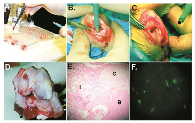
(a–c) Fresh aspirates of bone marrow were mixed with 1010 vp of Ad.Luc or Ad.GFP, and allowed to coagulate in individual wells of a 96-well plate. The clots were removed with forceps, loaded into a 3 mm diameter tube fitted with a plunger, and implanted into 3 × 8 mm osteochondral defects generated in the femoral condyles of rabbits. At periodic intervals postimplantation, the knees were harvested and implants examined. (d and e) Example of a clot implant at day 7; the clots were maintained within the lesion, and had integrated into surrounding tissues. (f) Fluorescent microscopy of the synovial lining of joints receiving clots seeded with Ad.GFP. There is little transduction of synovial cells.
From reference 11, with permission.