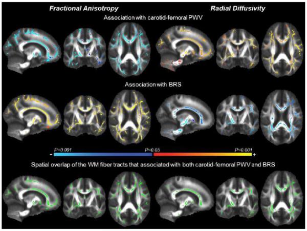
Figure 1.
Tract-based spatial statistic (TBSS) maps exhibit white matter (WM) fiber tracts with fractional anisotropy (left) and radial diffusivity (right) that associated with carotid-femoral pulse wave velocity (PWV) (top) and baroreflex sensitivity (BRS) (middle). The color bar illustrates the directionality and P-value of the associations. The bottom images show spatial overlap of the WM fiber tracts (green) that associated with both carotid-femoral PWV and BRS.

