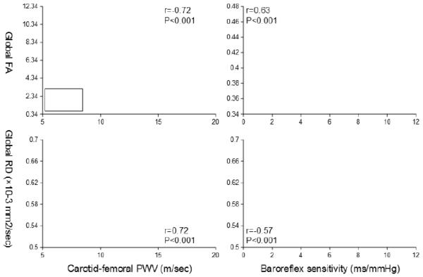
Figure 3.
Scatter plots show simple correlations of carotid-femoral pulse wave velocity (PWV) (left) and baroreflex sensitivity (right) with fractional anisotropy (FA) (top) and radial diffusivity (RD) (bottom). Mean values of FA and RD were extracted from the global WM skeleton that associated with both carotid-femoral PWV and baroreflex sensitivity (see the bottom of Figure 1).

