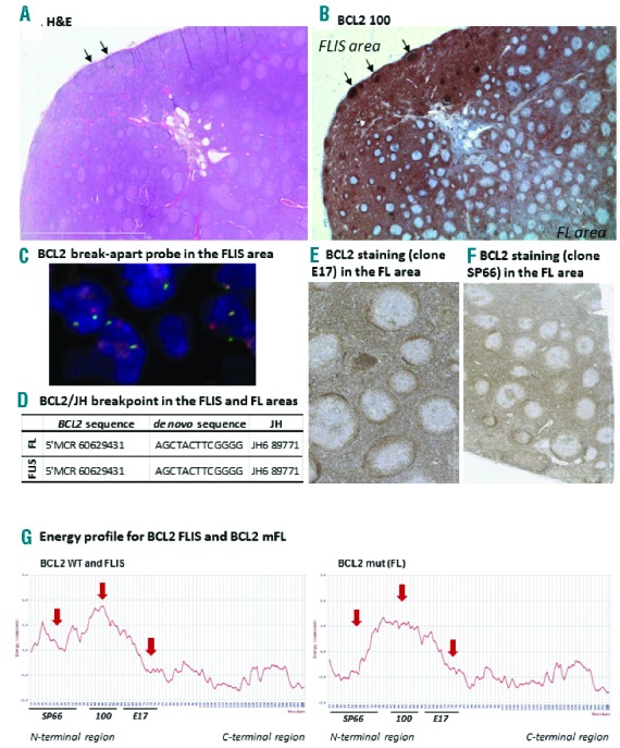
Figure 1.

Description of BCL2 status in both the FLIS and the FL areas of a cervical lymph node. (A) Hematoxylin/eosin coloration showing the FLIS and FL zones. (B) Histochemical staining of BCL2 with the E100 clone. The staining was negative in the germinal center of the FL areas, whereas it was extremely intense within the GC of the FLIS containing area (stronger than BCL2+ cells of the extra follicular zones) (C) FISH staining with the BCL2 break apart probe (LSI BCL2 break-apart probe, Vysis®) in the FLIS area. Similar results were obtained in the FL area. (D) Sanger sequencing of the BCL2/JH breakpoint, and the de novo inserted sequence, in the FLIS and FL areas. (E) Histochemical staining of BCL2 with the E17 clone in the FL area (F) Histochemical staining of BCL2 with the SP66 clone in the FL area. (G) Energy profile obtained from the BCL2 sequence obtained from the FLIS and the FL. Fixation sites of the 3 tested antibodies are mentioned.
