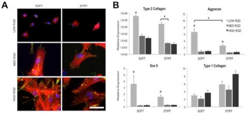
Figure 6. Scaffold mediated instruction upon arrival.
A) Vinculin localization (green), actin cytoskeleton (red), and nuclei (blue) staining of human mesenchymal stem cells (MSCs) cultured for 24 hours on fibrous HA scaffolds with varied RGD density and fiber modulus. Scale bar: 50 μm. B) MSC expression of chondrogenic markers after 14 days of culture in chondrogenic medium on fibrous HA scaffolds with varied RGD density and fiber modulus. *denotes significance (p<0.05) between groups, and  denotes significance (p<0.05) compared to other RGD densities within the same fiber stiffness condition. Adapted with permission from [11].

