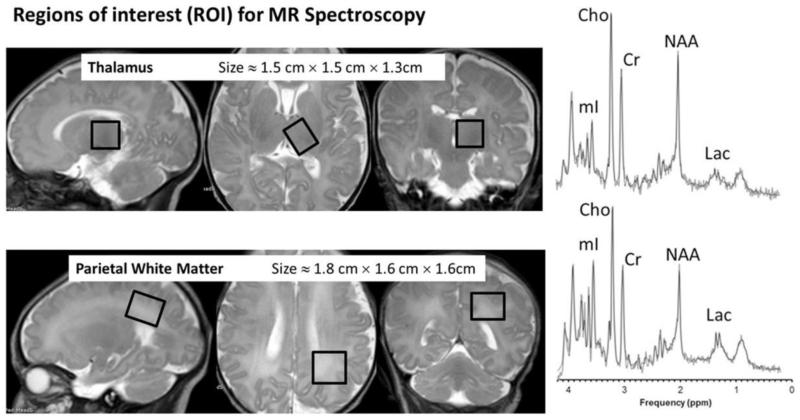
Fig 2. Short echo MR spectroscopy in Newborn Brain.
T2-weighted sagittal, axial, and coronal MR images of the newborn brain are shown above. The boxes indicate the region of interest (ROI) from where MR spectra will be acquired, with their approximate dimensions provided. The top row of images shows sagittal, axial and coronal images through the left thalamus, with an ROI in the thalamus. At the end of the first row, the spectrum appears normal with no associated abnormality. The bottom row of images shows sagittal, axial and coronal images through the left parietal lobe, with an ROI in the parietal white matter. The spectrum at the end of the row shows an increase in the lactate peak, compatible with hypoxic ischemic injury (HIE). Note that the both ROIs are drawn slightly oblique in order to maximize the sampled volume in the area of interest and avoid partial volume effects. (spectra courtesy of Dr. Stefan Bluml)

