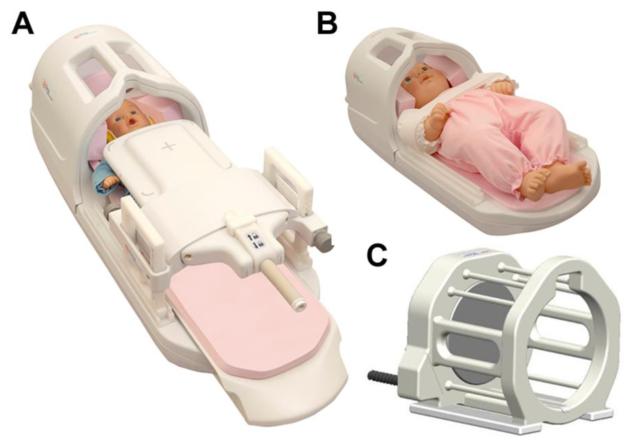
Fig 5. Neonatal Coils.
Images of three custom neonatal and infant sized head coils, which can be used to improve signal to noise ratio on examination. (A) Infant Cocoon, which can be used to image infants from 0-6 months of age. (B) Infant Head Spine Array, which can be used to image infants 0-6 months of age. (C) Neonatal Head Coil, which can be used in infants 0-1 months of age. The above coils can all be integrated with the neonatal incubator show in Fig 4. (coil images courtesy of Ravi Srinivasan)

