Fig. 3.
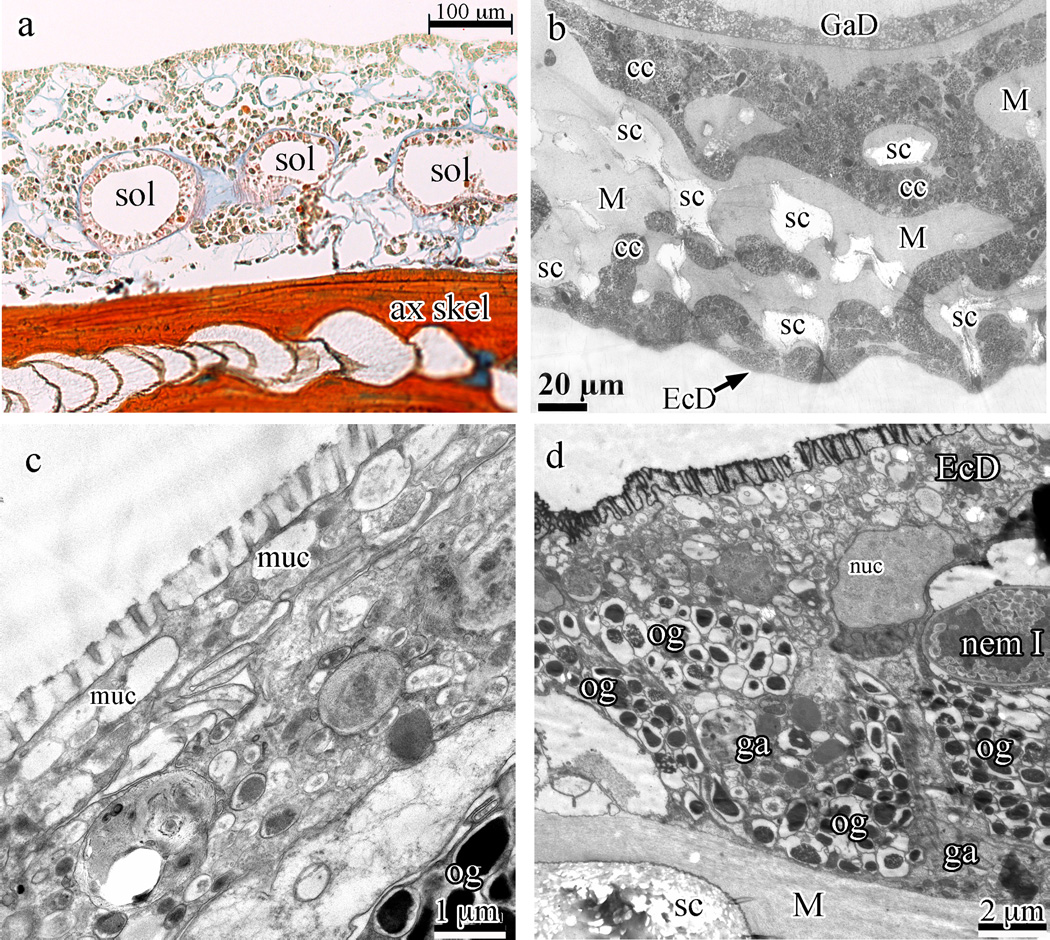
Swiftia exserta coenenchyme and ectoderm. (a) Longitudinal paraffin section stained with Mallory’s aniline blue (connective tissue stain) showing the hollow axial skeleton (ax skel) at the bottom, several circular solenia (sol) in the mesoglea between the skeleton and the outer rind of cells, and the blue mesoglea. (b) A cross-section of the coenenchyme by TEM shows light areas within the coenenchyme that hold remnants of sclerites (sc) which do not stain with electron-dense dyes. Ectoderm (EcD) is found at the bottom of the image and gastroderm (GaD) at the top. Cell cords (cc) can be seen permeating the fibrous mesoglea (M). (c and d) High magnification TEM shows the microvilli in section and the thin, flattened ectoderm cells filled with electron-lucent granules (panel c) while panel (d) gives a survey of the stratified rind of the coenenchyme, with thin ectoderm cells (EcD) at the top covering oblong granular cells with electron-dense granules and amorphously shaped granular amoebocytes. At center-right is a type I nematocyst (nem I). Oblong granular cells (og), granular amoebocytes (ga), fibrous mesoglea (M), and sclerites (sc) are indicated in the panels.

