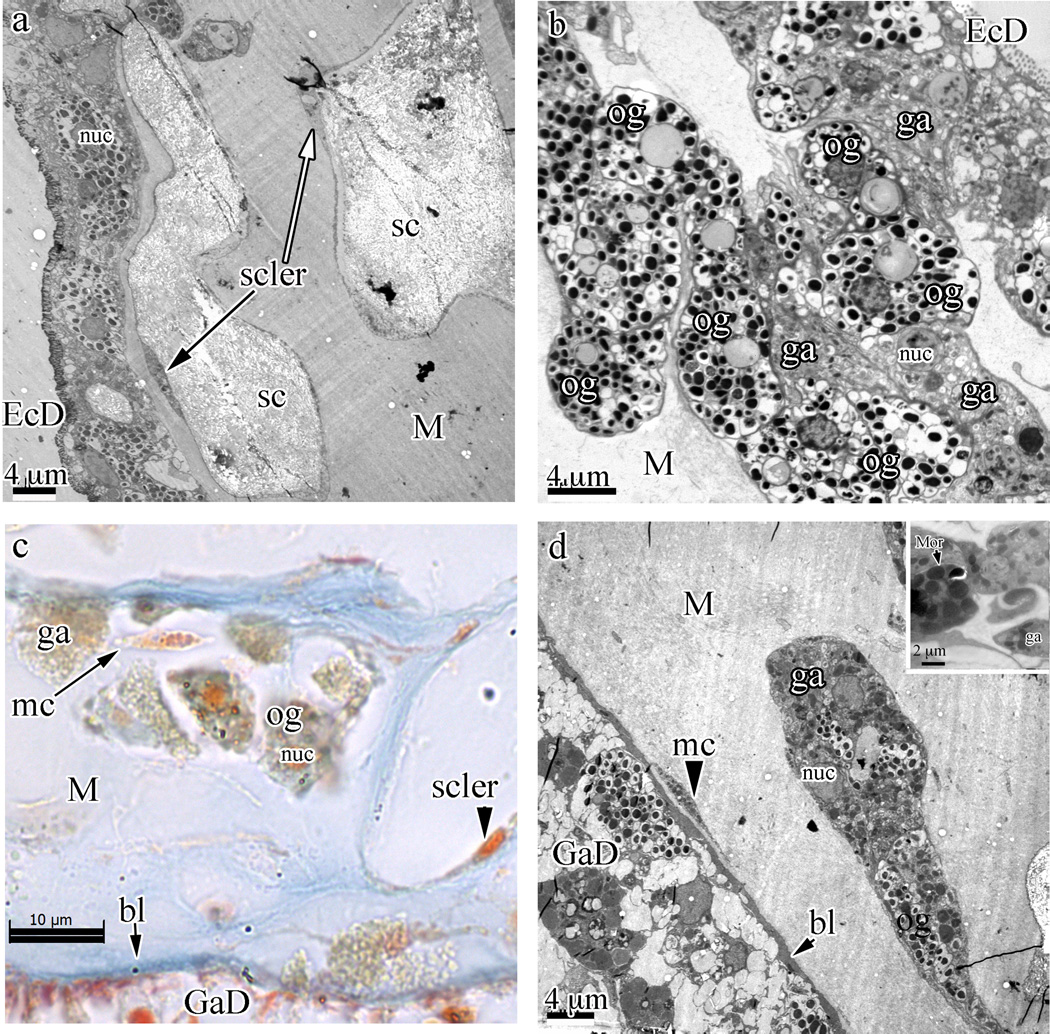
Fig. 4.
Cell cords in the coenenchyme of Swiftia exserta with embedded sclerites (sc). (a) TEM overview showing the cellular architecture of the coenenchyme from the ectoderm (EcD) into the fibrous mesoglea (M). (b) A closer view of the dense packing and the irregular shape of the cell cords filled with oblong granular cells (og, with electron-dense granules) and granular amoebocytes (ga, of indeterminate shape and with granules staining less intensely and of different sizes) shows the continuity of cells from beneath the thin ectodermal cells into the cell cords. (c) Paraffin section stained with Mallory’s aniline blue showing the acellular nature of the mesogleal matrix. The orange nucleus of a sclerocyte is visible towards the bottom right of the sclerite space (small arrowhead). Blue staining of the fibrous mesoglea matrix indicates a collagen-like stain affinity. An isolated, single mesogleal cell (mc) is indicated by a long arrow. The basal lamina (bl) is indicated by a short arrow. (d) An area of the mesoglea with an isolated cell cord embedded within the fibrous mesogleal matrix shows a single amoebocytic mesogleal cell (arrowhead to mc) and the basal lamina (short arrow to bl) that separates the gastroderm cells (GaD) from the mesoglea (M). The insert at the top right shows a morula-like cell (Mor).

