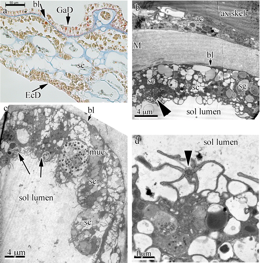
Fig. 5.
Solenia-type endo-/gastroderm in Swiftia exserta. (a) Solenia-type gastroderm (GaD) lining is visible at the upper part in a paraffin section stained with Mallory’s stain. The staining indicates that one of the digestive cell types is acidophilic (bright orange-red granules in cytoplasm). A basal lamina (bl) structure isolates the gastroderm from the mesoglea. (b–d) Transmission electron micrographs of solenia show different aspects. A solenium near the axial skeleton (ax skel) and axial epithelia (ae) is seen. This solenium is separated from the fibrous mesoglea (M) by a basal lamina-like layer (short arrow to bl). This basal lamina layer is faintly visible in panels (b) and (c), and clearly visible in panel (a) (arrow and bl). The arrowhead indicates a zymogen-like cell. Several secretory cells (sc) and a secretory granule (sg) are indicated. (c) Low magnification TEM of a section through a solenia (sol) showing several types of digestive cells. Long arrows indicate phagocytic vesicles. Short arrow indicates the basal lamina-like structure. A mucus-secreting cell’s granules are indicated (muc), as are several secretory cells (sc). Panels (b) and (c) show secretory cells (sc) packed with granules (sg) and phagocytic cells with membrane ruffles. To move liquid through the solenia (sol lumen), a flagellated cell with flagellum, seen in panel (d) with a section perpendicular to the flagellum (arrowhead) and base, is used.

