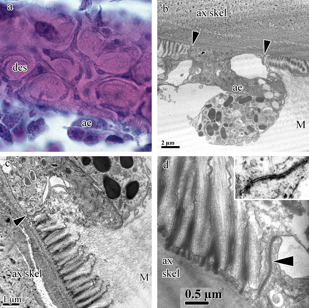
Fig. 6.
Swiftia exserta desmocytes and axial epithelia. (a) Desmocyte plaques (des) and axial epithelia (ae) with H&E staining from paraffin sections. (b–d) TEM images demonstrate the elevation of the axial epithelial cells (ae) above the plane of the desmocytes (panel b) as well as an active axial epithelial cell capable of secreting gorgonin. Panels (c) and (d) show the fine structure of the desmocyte anchors into the mesoglea and the many secretory vesicles and golgi bodies found in the axial epithelial cells (panel c). Insert in (d) shows the septae found in these desmocyte-to-axial epithelium junctions. Junctions between the desmocyte and axial epithelium cell are indicated by arrowheads in panels (b–d). The axial skeleton (ax skel) and mesoglea (M) are indicated in panels (b–d).

