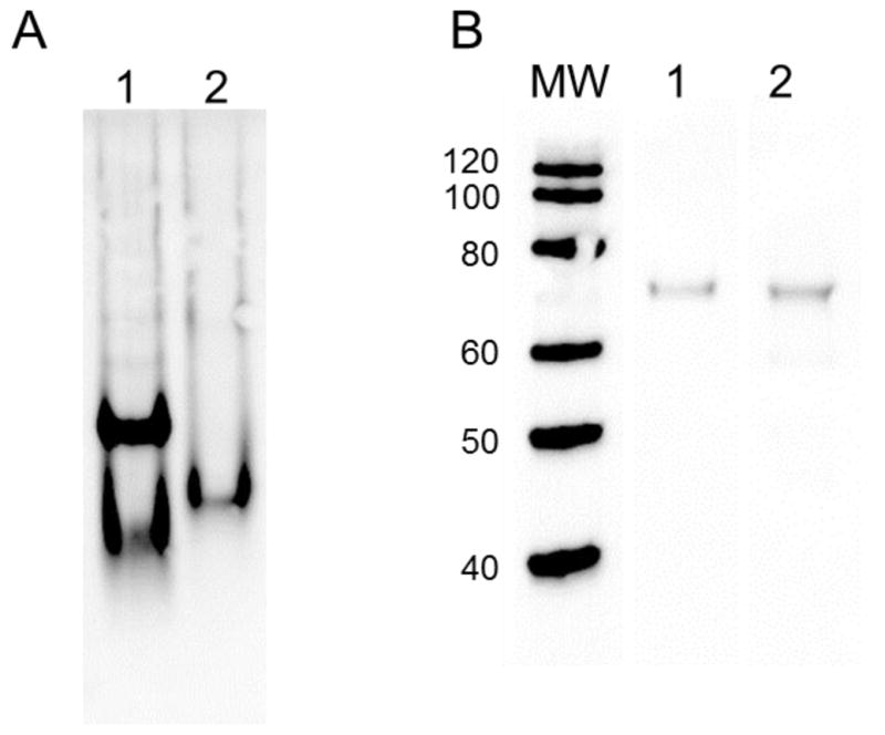
Fig. 3.

Western blot analysis of PAFAH-II constructs resolved by native PAGE and SDS PAGE. (A) Western blot analysis resolved by native PAGE: lane 1: WT-PAFAH-II-YFP-His, lane 2: G2A mutant-PAFAH-II-YFP-His, and blotted with GFP specific antibodies. WT-PAFAH-II displays monomer and dimer bands, while the myristoyl mutant resolves as a monomer. (B) Western blot analysis resolved by SDS PAGE: molecular weight marker lane (kDa) on left, lane 1: WT-PAFAH-II-YFP-His, lane 2: G2A-PAFAH-II-YFP-His, and blotted with GFP specific antibodies. The wild type and G2A-PAFAH-II fusions with YFP run at their expected molecular weight of 77 kDa.
