Figure 1.

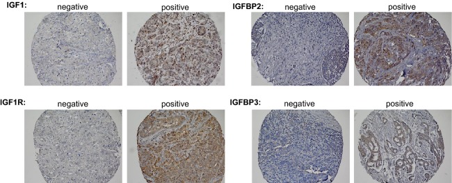
Immunohistochemical staining of IGF-axis proteins in breast cancer tissue. Negative and positive staining for IGF1, IGF1R, IGFBP2, and IGFBP3 expression. Individual tissue cores at 20× magnification.

Immunohistochemical staining of IGF-axis proteins in breast cancer tissue. Negative and positive staining for IGF1, IGF1R, IGFBP2, and IGFBP3 expression. Individual tissue cores at 20× magnification.