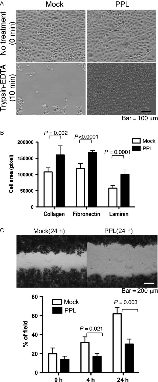
Figure 3.

Forced PPL expression promoted cell adhesion. (A) Cell detachment assay. The same numbers of mock-transfected or PPL-transfected KYSE270 cells were cultured in 10-cm dishes for 24 h and then treated with 1 mL trypsin-EDTA for 10 min no treatment, 0 min. (B) Cell adhesion assay. Mock-transfected or PPL-transfected KYSE270 cells were fluorescent-labeled, and incubated for 1 h in plates coated with the indicated extracellular matrix. The area of adherent cells was measured. Two random fields were counted in three separate wells, and the results were shown as mean + SD. (C) Migration assay. Mock-transfected or PPL-transfected KYSE270 cells were grown to confluence in a 24-well plate with an insertion to keep a cell-free area. After the removal of the insertion, cells were cultured for indicated times and images were captured. Cell migration was quantified by measuring the % of area covered with cells. Data are shown as mean + SD of triplicate assays. PPL, periplakin.
