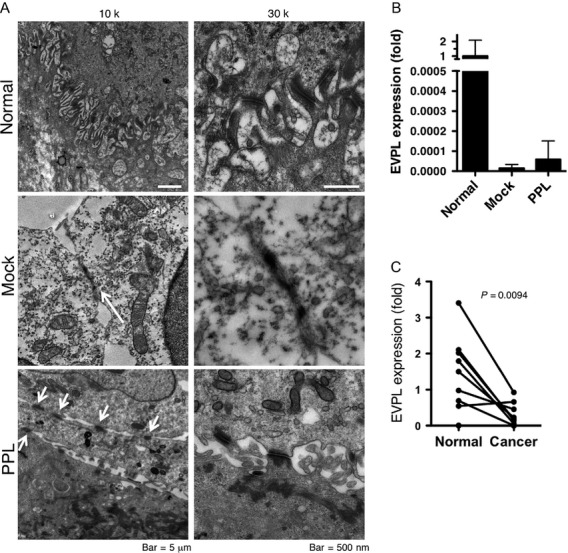
Figure 4.

Forced PPL expression induced desmosome-like structures. (A) TEM images were obtained from normal human esophageal mucosa (normal) and mock-transfected or PPL-transfected KTSE270 cells at 10,000× or 30,000× magnification. Desmosomes were frequently found in normal tissues and PPL-transfected cells (arrows). In the images of the mock-transfected cells taken at 10,000× magnification, the arrow indicates adhesion plaque-like structures, which was not identified as a desmosome at higher magnification. (B) Expression of envoplakin (EVPL) in mock- or PPL- transfected KYSE270 cells. EVPL mRNA levels were shown as fold expression of the levels to normal esophageal mucosa (average of 13 mucosa = 1). Data are shown as mean + SD of three assays. (C) Expression of envoplakin (EVPL) in ESCC tissues with paired normal mucosa. PPL, periplakin; ESCC, esophageal squamous cell carcinoma.
