
Figure 4.
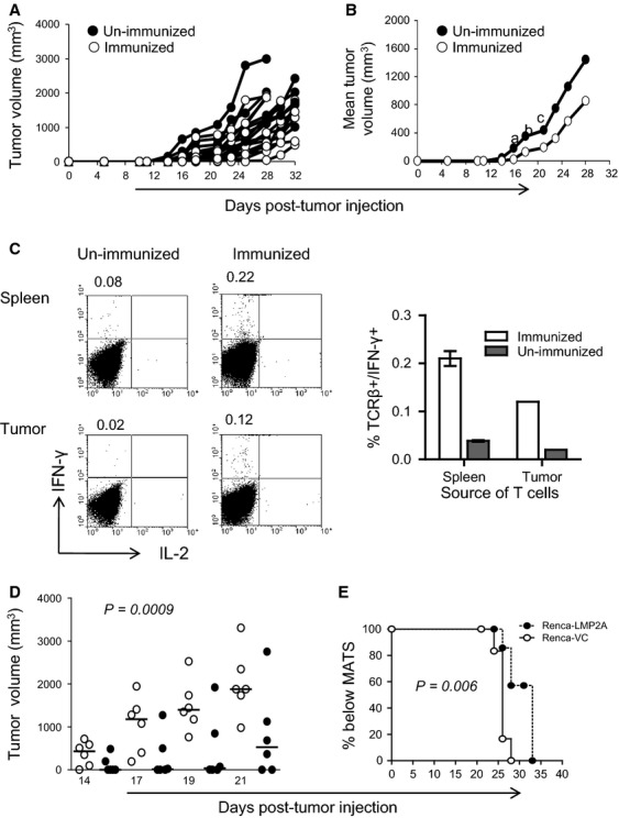
Control of Renca-LMP2A tumor growth in vivo by Vac-LMP2A. On day 0 BALB/c mice were immunized with recombinant Vac-LMP2A (n = 10) or unimmunized (n = 10) 11 days later, all mice from both groups were injected with Renca-LMP2A, and tumor growth measured up until day 32. Graphs depict (A) individual and (B) mean Renca-LMP2A tumor growth among unimmunized (•), and Vac-LMP2A-immunized mice (○). Two-tailed Mann–Whitney nonparametric test was used to compute the P-values for: a day 14; P = 0.007, b day 16; P = 0.004, and cday18; P = 0.02. (C) At the end of the tumor-monitoring period, cells were isolated from the spleens and tumors from three mice in each group and used to set up T-cell cultures using irradiated Renca-LMP2A as APCs. After three successive weekly restimulations, cultures were tested for IFN-γ and IL-2 production by ICS. Representative FACS plots show percentage of IFN-γ versus IL-2 among splenocytes and the bar graph shows the mean ± SEM of IFN-γ+ TCRβ+ cells among cultured splenocytes (spleen, n = 3) or TIL (pooled n = 3 tumors) from immunized or unimmunized animals. Numbers at the top of the FACS plots indicate the percentage of IFN-γ+ cells. D&E Mice immunized with Vac-LMP2A were challenged on day 11 with either Renca-VC (•) or Renca-LMP2A (○) and tumor growth monitored periodically as a surrogate for immune control. (D) Scatter plot showing tumor volumes of individual mice. Horizontal lines indicate the median. One-way ANOVA and nonparametric analysis was used to compute the P-value. (E) Top rank analysis of time taken to reach the MATS.