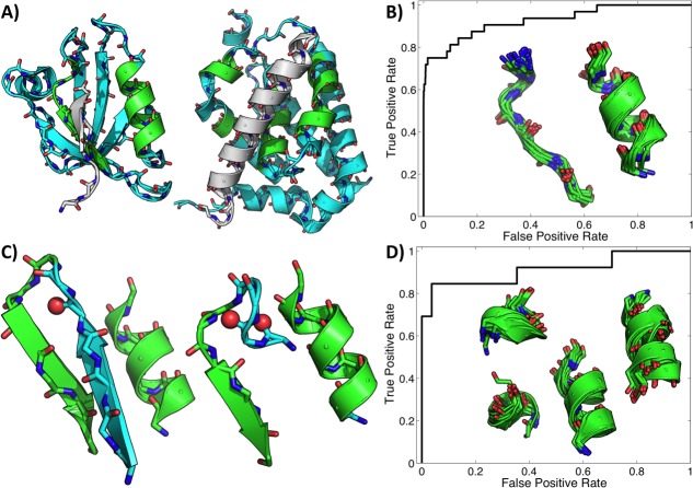
Figure 5.
Simple binding site fragments from single representatives of PDZ and Bcl-2 family members serve as signatures of function. (A) Shown are a PDZ domain (first PDZ domain of MAGI-1, PDB ID 2I04; left) and a Bcl-2 family member (Bcl-2-like protein 10, PDB ID 4B4S; right) bound to their cognate peptides (gray). The regions excised as potentially representative of the binding function are shown in green. Receiver operating characteristic curves of RMSD to the excised motifs as classifiers of family membership for PDZ and Bcl-2 domains are shown in (B) and (D), respectively, along with a superposition of close hits. Only matches below 2.0 Å were counted as predictions, and this set included all true domains in both cases. Shown in (C) are two common modes of protecting the PDZ binding loop by engaging the otherwise exposed amides in internal hydrogen bonds.

