Figure 2.
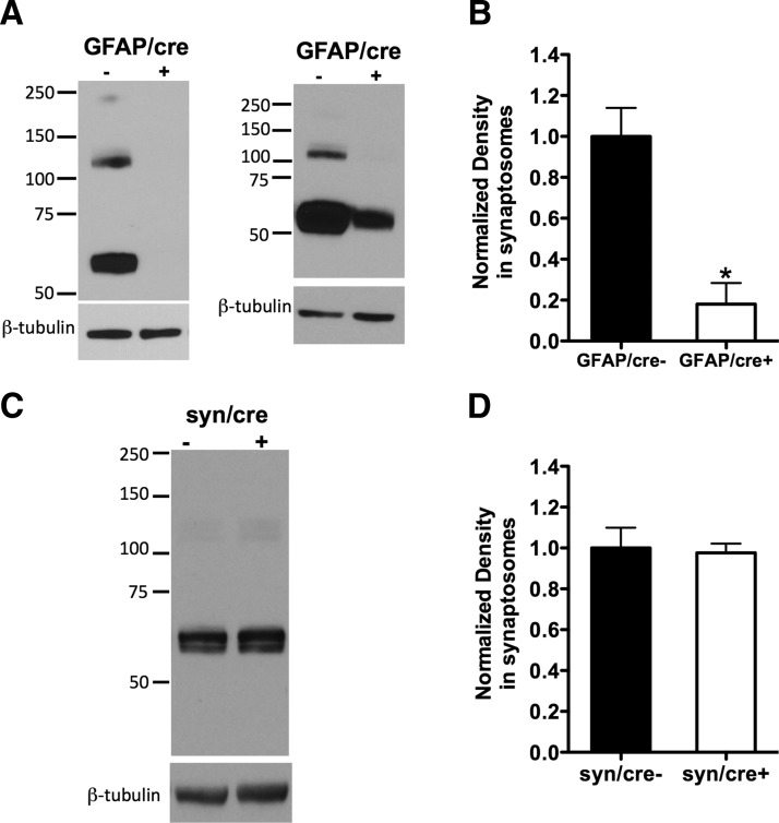
The Cre-driver GFAP produces great loss of GLT-1 expression in conditional GLT-1 knock-out mice, whereas synapsin-Cre produces no detectable loss of GLT-1 protein. A, Protein immunoblot analysis using anti-nGLT-1 antibody in crude synaptosomes isolated from GLT-1Δ/Δ;GFAP-Cre ERT2 mice (referred to as GFAP/cre+) and their littermate controls (GFAP/cre−): in two GFAP/cre+ mice, the GLT-1 protein was almost completely knocked out (left blot); in another two GFAP/cre+ mice, the GLT-1 protein was greatly but not completely (∼35% remaining) deleted (right blot). B, Pooling densitometry results from the four experiments that were performed, there was an ∼80% reduction of GLT-1 expression (*p < 0.05) in GFAP/cre+ (n = 4) mice compared with controls (n = 4). C, Immunoblot assay of GLT-1 expression in GLT-1Δ/Δ;Syn-Cre mice (referred to as syn/cre+) compared with control mice (syn/cre−). D, Densitometry analysis of GLT-1 expression in syn/cre+ and syn/cre− mice revealed no change (p > 0.05, n = 3). GLT-1 monomer is at ∼60 kDa (dimer at ∼120 kDa and multimer at ∼250 kDa). All GLT-1 forms (if present) were included in the densitometry analysis.

