
Figure 2.
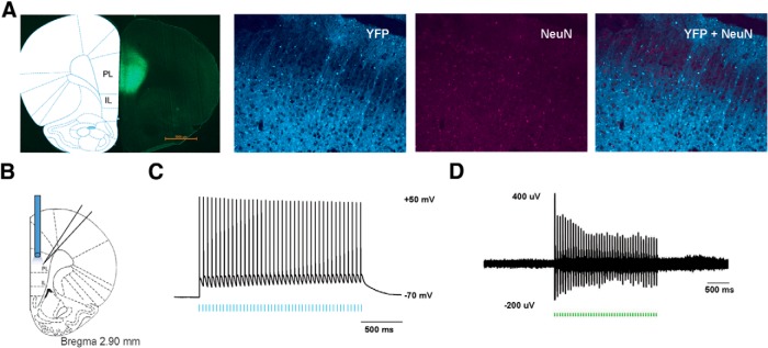
Functional expression of ChR2 in PL-PFC neurons. A, Histologic expression of ChR2-eYFP in the PL-PFC 14 d after viral injection. ChR2-eYFP was found to be restricted to neurons in the PL-PFC region. NeuN, Neuronal marker. B, Schematic showing electrophysiological recordings were conducted at the site of light delivery. Data were collected from prefrontal neurons 14 d after viral injections in the PL-PFC. C, Whole-cell patch-clamp recording showed that direct illumination with an LED in brain slices containing the PL-PFC elicited spikes with high fidelity in prefrontal neurons. LED pulses are represented below in blue. D, In vivo recording at the virus injection site with simultaneous photoactivation demonstrated that laser pulses reliably triggered spikes in PL-PFC neurons. Laser pulses are represented below in green.