Figure 4.
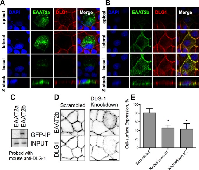
Localization of the PDZ protein DLG1 and EAAT2b in MDCK cells. A, Stable expression of EGFP-EAAT2a in MDCK cells. EAAT2a expression (green, second column) demonstrates apical (top) and basolateral expression of the carrier throughout the cell (bottom two panels). B, EGFP-EAAT2b expression in MDCK cells, however, is restricted to the basolateral surfaces. Similar localization is seen for DLG1 at the basolateral membranes (red). C, Immunoblot of GFP fusion proteins immunoisolated from either the stable EGFP-EAAT2a or EGFP-EAAT2b cell line. DLG1 was only found with the EGFP-EAAT2b protein. D, Cross sections of MDCK cells expressing EGFP-EAAT2b. A scrambled shRNA (left) has no effect on the surface expression of EAAT2b (top) or the expression of DLG1 (bottom). Knockdown of DLG1 (right) is apparent in one cell in the center of the field of view (bottom). That cell has an increase in cytosolic localization of the EGFP-EAAT2b protein (top). Scale bars, 50 μm. E, Quantitation of two different DLG1 knockdown constructs reveals a loss of surface expression of EAAT2b. *p < 0.0001 by one-way ANOVA (n = 9). Error bars indicate SEM.

