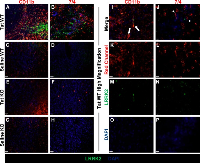
Figure 8.
LRRK2 is not expressed in neutrophils in the CNS of Tat-injected mice. A–H, IHC for CD11b (red, left column), 7/4 (red, right column), LRRK2 (green), and DAPI (blue) in WT Tat-injected animals (A, B), WT saline-injected animals (C, D), LRRK2 KO Tat-injected animals (E, F), and LRRK2 KO saline-injected animals (G, H) (20×). I–P, IHC for CD11b (red, left column), 7/4 (red, right column), LRRK2 (green), and DAPI (blue) in high-magnification (60×) merge (I, J) and single-channel images (K–P) for WT Tat-injected animals (I, white arrows depict CD11b- and LRRK2-positive cells; J, white arrowheads depict 7/4-postive LRRK2-negative cells). Scale bars: A–H, 32 μm; I–P, 11 μm.

