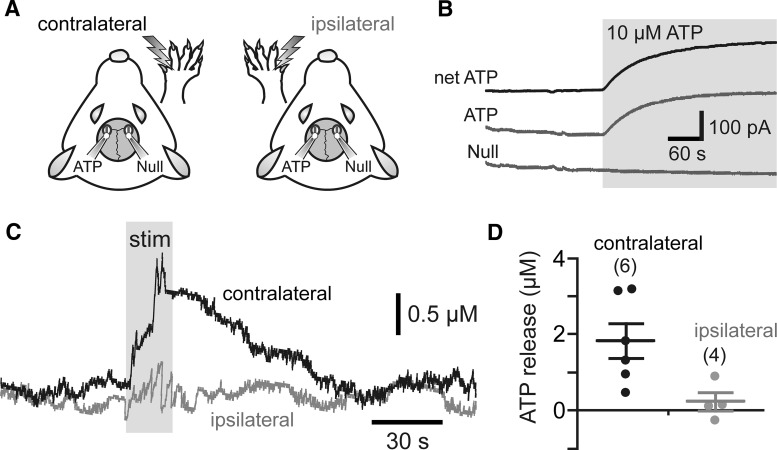
Figure 1.
Activation of somatosensory pathways increases extracellular concentration of ATP in the cerebral cortex of an anesthetized rat. A, Schematic drawing of the experimental design illustrating positioning of the ATP or null biosensors on the exposed surface of the forepaw region of the SSFP. B, Calibration of biosensors illustrating changes in ATP and null sensor currents in response to ATP (10 μm). To determine changes in ATP concentration, null sensor currents were subtracted from ATP sensor currents (net-ATP). C, Representative recordings showing changes in ATP concentration within the SSFP region in response to electrical stimulation of the contralateral and ipsilateral paws. D, Summary data illustrating peak ATP release triggered in the SSFP region by electrical stimulation of the contralateral (n = 6) and the ipsilateral (n = 4) paws.

