
Figure 5.
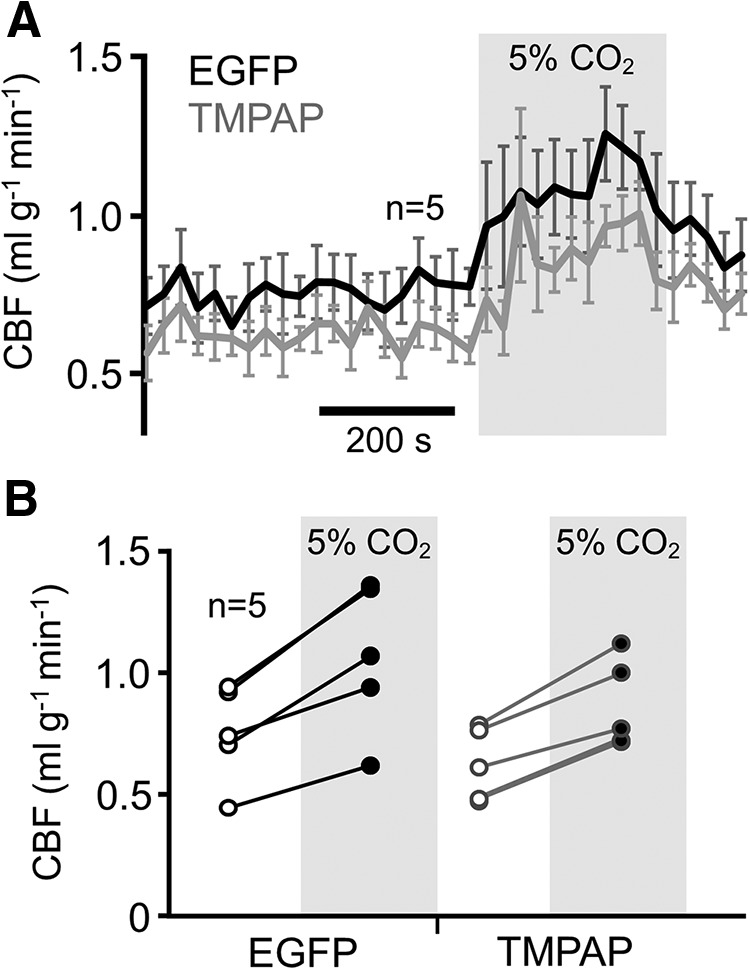
TMPAP activity in the SSFP region does not affect local CBF and cerebrovascular reactivity. A, Mean time course of the CBF determined using arterial spin labeling MRI within the SSFP regions transduced to express EGFP (black) and TMPAP (gray) at resting conditions and in response to CO2 challenge (5% inspired CO2). B, Summary data illustrating resting CBF and average increases in CBF in response to CO2 in the SSFP regions expressing EGFP and TMPAP.