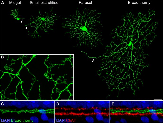
Figure 1.
Morphology of broad thorny ganglion cells. A, An ON midget, a small bistratified, an ON parasol, and a broad thorny ganglion cell are shown. Cells were injected with Lucifer yellow at similar retinal eccentricities. Maximum intensity projections of confocal image stacks were separately produced and assembled for this image. Arrowheads point to the axons of the cells. B, Magnification of a broad thorny cell's dendritic tree (z-projection). C–E, Lucifer-yellow-injected broad thorny cells were counterstained in whole-mounted tissue with DAPI to label cell nuclei and antibodies against ChAT to label starburst amacrine cells. An xy-projection of a magnified area from a broad thorny cell's dendritic field is shown. Scale bars: A, 50 μm; B, E, 10 μm.

