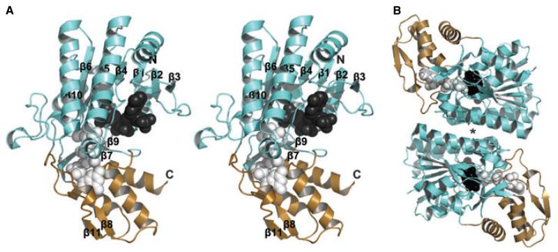
Fig. 9.
Structure of RMD from A. thermoaerophilus. (A) Stereoview of the RMD monomer. The cofactor-binding domain and the substrate-binding domain are shown in aqua and light sand, respectively; the APPR portion of the cofactor (dark gray) and the ligand analog GDP-d-Man (light gray) are represented as space-filling models. Termini and secondary structural elements are labeled. (B) View of the RMD homodimeric structure; an asterisk highlights the four-helix bundle, the typical SDR enzyme dimerization mode.

