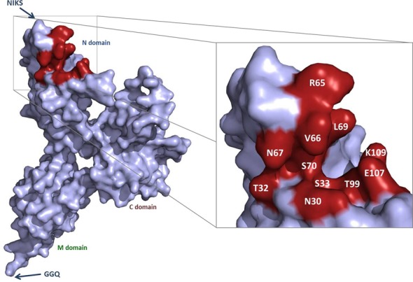
Figure 1.

P1 pocket residues mutated to alanine. Representation of the structure of eRF1, with the three domains and the conserved NIKS and GGQ motifs indicated. The magnified view shows the surface of the P1 pocket. The residues mutated to alanines are shown in red.
