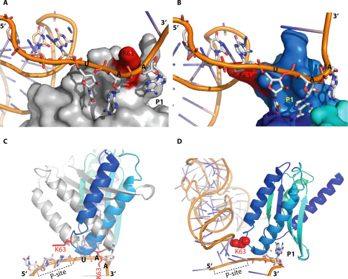
Figure 5.

Interactions between the stop codon and the eRF1 N-terminal domain. (A) The first step in the recognition process, with the stop codon located near the NIKS motif (K63 in red) and the P-site tRNA shown in orange on the left. (B) Second step in the recognition process, involving the interaction of the stop codon with the P1 pocket. The P-site tRNA is shown in orange on the left. (C) The interaction between the stop codon and the P1 pocket requires changes in the conformation of the N domain. eRF1 is shown in grey for the conformation adopted during the first step, as in panel (A), and in blue for the conformation adopted during the second step. The P-site tRNA is not shown, for the sake of clarity. (D) As in panel (B), eRF1 is in the conformation required for the second step of stop codon recognition, with K63 indicated in red.
