
Figure 4.
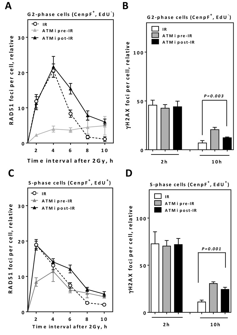
ATM inhibition after end resection results in higher numbers of residual RAD51/γH2AX foci in both S and G2 cells. (A) A549 cells were EdU pulse labeled, irradiated with 2 Gy and the kinetics of RAD51 foci numbers were monitored in CenpF+/EdU− G2-cells at the indicated time points. (B) γH2AX foci were evaluated at the indicated time points in CenpF+/EdU− G2-cells. (C) The kinetics of RAD51 foci numbers were monitored in CenpF+/EdU+ S-cells at the indicated time points. (D) γH2AX foci were evaluated in CenpF+/EdU+ S-cells at the indicated time points. In all cases, the number of foci measured in non-irradiated cells was subtracted from that observed in irradiated cells. Error bars represent the SEM of three independent experiments.