
Figure 4.
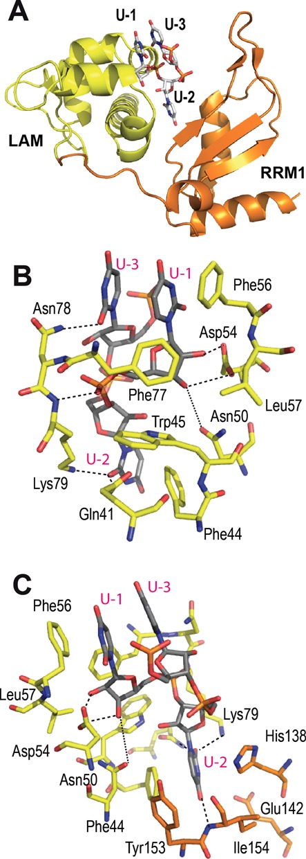
Binding site of the terminal uridines in LARP7. (A) LARP7 La module with LAM domain in yellow and RRM domain in orange, showing the arrangement of the terminal uridines. (B) Details of the 3′ uridines binding site viewed from the LAM side, showing the terminal ribose binding Asn50 and Asp54 and the stacking of U-1 and U-3 with Phe56, in stick representation, with carbons gray for RNA, yellow for amino acids from LAM. Dotted lines indicate H-bonds (distances in the range of 3.0–4.0 Å). (C) Perpendicular view, turned around the vertical axis, showing the specific recognition of U-2 by the RRM1, in orange.