
Figure 10.
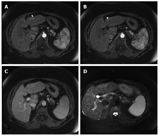
Arterioportal shunt. Post-contrast fat-suppressed 3D-GRE T1-weighted images during the late hepatic arterial (A and B) and delayed phases (C); D: Fat-suppressed SSFSE T2-weighted image. There is a convoluted linear area of increased arterial enhancement with a vessel leading to it (arrowhead, A, B), which does not demonstrate delayed washout (C), or corresponding T2 signal abnormality in keeping with an AP shunt. SSFSE: Single-shot fast spin-echo; GRE: Gradient recalled echo.