
Figure 8.
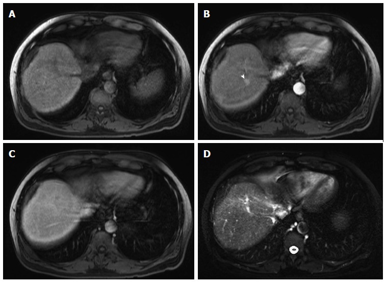
High-grade dysplastic nodules. Pre- (A) and post-contrast fat-suppressed 3D-GRE T1-weighted images during the (B) Late hepatic arterial; and (C) Delayed phases; (D) Fat-suppressed SSFSE T2-weighted image. There is a nodule at hepatic segment #7, which demonstrates iso T1 signal intensity (A), increased arterial enhancement (arrowhead, B), without definite washout or corresponding T2 signal abnormality in keeping with a HGDN. However, early HCC cannot be totally excluded and short-term follow-up should be recommended, especially in patients with hepatitis C infection. HGDN: High-grade dysplastic nodules; HCC: Hepatocellular carcinoma; GRE: Gradient recalled echo; SSFSE: Single-shot fast spin-echo.