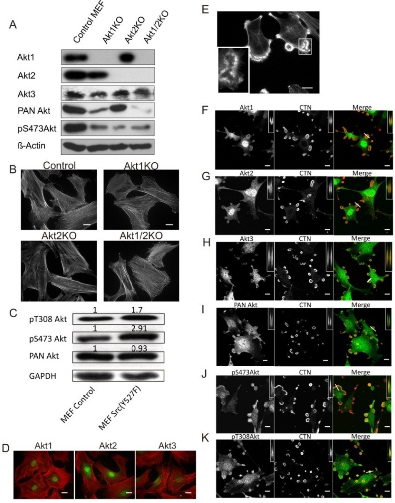
Figure 1.
Src Enhances Akt Phosphorylation and Its Localization to Podosomes. (A) Western blots showing the Akt isoform expression profile of Akt1KO, Akt2KO and Akt1/2KO MEF cells. β-Actin was used as a loading control; (B) Knockout of Akt1 and/or Akt2 does not affect cell morphology. Akt1KO, Akt2KO and Akt1/2KO MEF cells were stained for F-Actin using TRITC-phalloidin. Scale bars represent 20 μm; (C) Src (Y527F) activates Akt. Whole cell lysates from control MEF cells and Src (Y527F) cells were analyzed by Western blots using antibodies against pT308 Akt or pS473 Akt, and PAN Akt. The numbers above the bands indicate staining intensity relative to control cells using β-Actin as a loading control; (D) Akt1, Akt2 and Akt3 are expressed in MEF cells showing prominent nuclear localization. Akt (green) was immune-stained with isoform-specific antibodies, and F-actin (red) was stained with TRTIC phalloidin. Scale bars represent 20 μm; (E) MEF cell expressing Src (Y527F) produces podosomes and rosettes stained with TRITC-phalloidin (F-actin). Outline in white indicates the area taken for the inset in the lower left corner showing that rosettes are composed of individual podosome dots which coalesce to form rosettes. Scale bar represents 20 μm; (F–K) Localization of Akt isoforms in Src-induced podosomes and rosettes. Confocal microscopic images of MEF cells stably expressing Src (Y527F) were immune-stained red for cortactin (CTN); green for Akt1, Akt2, Akt3, PAN Akt, pS473 Akt, and pY308 Akt. Insets are x-z axis scans of the plane represented by the line drawn through a selected rosette shown in the merged images, where Akt can been seen to localize to the edges of the rosette. Scale bars represent 20 μm.

