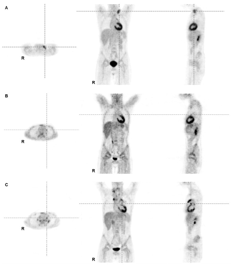
Figure 1.
FDG-PET images of an 11-year-old male patient with mixed cellularity HL stage II at staging (A), interim (B) and at restaging due to suspected relapse 16 months after initial diagnosis (C). (A) Lymphoma lesions at staging were found at left supra- and infraclavicular sites and in the mediastinum. The patient was treated according to the TG1 protocol (GPOH-HD2002P); (B) The interim PET demonstrated minimal residual uptake in the left mediastinum adjacent to the left atrium/ventricle primarily interpreted as physiological myocardial uptake (with no anatomical equivalent in the corresponding low-dose CT). This lesion was missed by the first truth-panel assessment (false negative by visual assessment). By semi-quantitative means the lesion exceeded SUV thresholds (true positive by semi-quantitative means); (C) PET at time point of restaging showed multiple areas of intense tracer uptake indicating a recurrence.

