Figure 8.
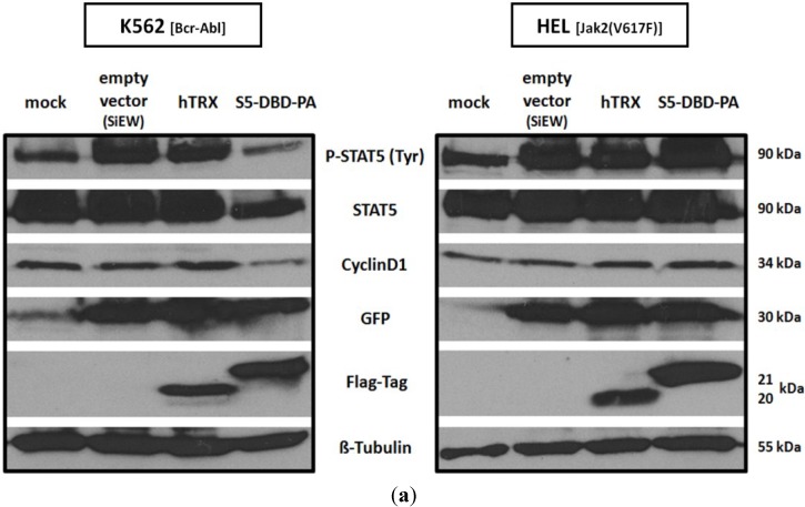
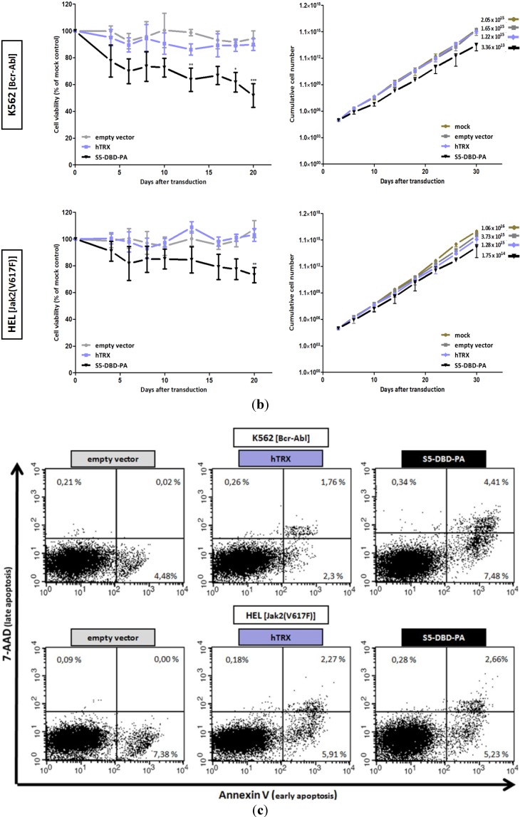
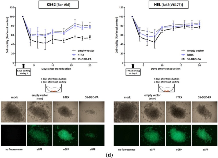
Reduction of K562 and HEL cell survival after infection with the S5-DBD-PA encoding lentivirus. (a) Bcr-Abl+ K562 and Jak2(V617F)+ HEL cells were infected with SiEW lentiviral vectors encoding either S5-DBD-PA, hTRX or an empty vector as controls. After 7 days, cell lysates were prepared and analyzed by western blotting for the expression of the encoded proteins with a Flag-tag antibody. Additional antibodies were used for the detection of Stat5 and P-Stat5 as well as for the analysis of eGFP fluorescence marker and Cyclin D1 target gene expression; (b) Proliferation and viability of the cells were monitored with the XTT-assay (n = 4; Ø ± SD), cell growth was analyzed by counting the cumulative cell numbers at each passaging interval (n = 3; Ø ± SD). Graphs indicate significantly reduced XTT-values (percentage of mock control) in comparison to empty vector expressing cells. * p < 0.05, ** p < 0.01, *** p < 0.001 (2-way-ANOVA with Bonferroni correction); (c) Analysis of apoptosis induction by Annexin V/7-AAD staining. 10 days after virus transduction cells were stained and analyzed by FACS. Divided FACS dot plots indicate unstained vital cells (lower left), early apoptotic cells positive for Annexin V (lower right), Annexin V/7-AAD double positive apoptotic cells (upper right) and late apoptotic/necrotic cells positive for 7-AAD (upper left); (d) eGFP expressing K562 and HEL cells were FACS sorted 2 days after infection with the lentiviruses and analyzed for changes in viability and growth by XTT conversion. Results are shown as the percentage of viable cells compared to mock control (n = 3; Ø ± SD). Significantly reduced XTT-values in comparison to empty vector expressing cells are indicated. ** p < 0.01 (2-way-ANOVA with Bonferroni correction). Phase contrast and fluorescence microscopy images of accumulated cells at the round-bottom of assay-96 well plates were taken 7 days after virus transduction (5 days after cell sorting and seeding).


