Fig. 1.
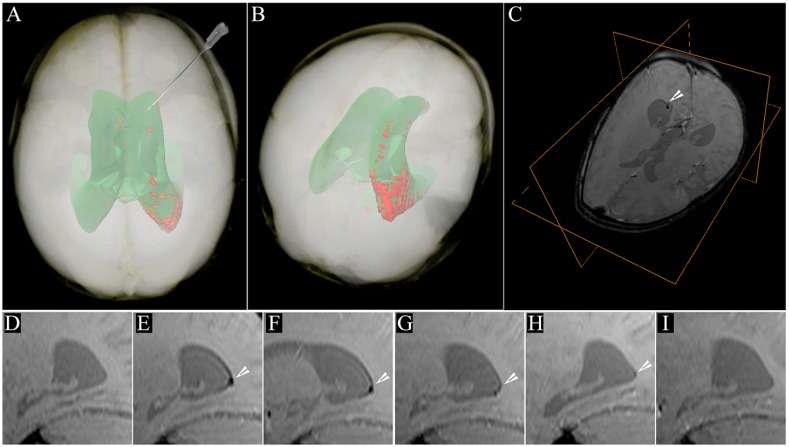
MRI of SPIO-labeled cells in a human brain. MRI evaluation of long-term tracking of transplanted cells labeled with superparamagnetic iron oxide (SPIO) nanoparticles in the human brain. SPIO-labeled autologous cord-blood-derived cells were transplanted in the frontal horn of the lateral ventricle of a patient with global cerebral ischemia. (A) 24 hours post-transplantation (PT) the distribution of the SPIO signal generated from the transplanted cells is detectable within the occipital horn of the right lateral ventricle (red); the projection of the ventricular system is shown in green. The needle indicates the route and trajectory of cell transplantation via the frontal horn. This is a volume rendering of MRI data of the patient’s head. (B) Location of the hypointense SPIO signal generated from transplanted cells in a postero-superior view of the patient’s head, where it can be better appreciated. (C) T2*-weighted image of the patient’s head with an orthogonal view centered on the cellular SPIO signal in the occipital horn (arrowhead). (D-I) The longitudinal dispersion of the cellular SPIO signal within the occipital horn (arrowheads) is shown in sagittal T2*-weighted MRI scans at different time points: (D) pre-transplantation; (E) 24 hours PT; (F) 7 days PT; (G) 2 months PT; (H) 4 months PT; and (I) 33 months PT. Figure and data reproduced with permission (Janowski et al., 2014).

