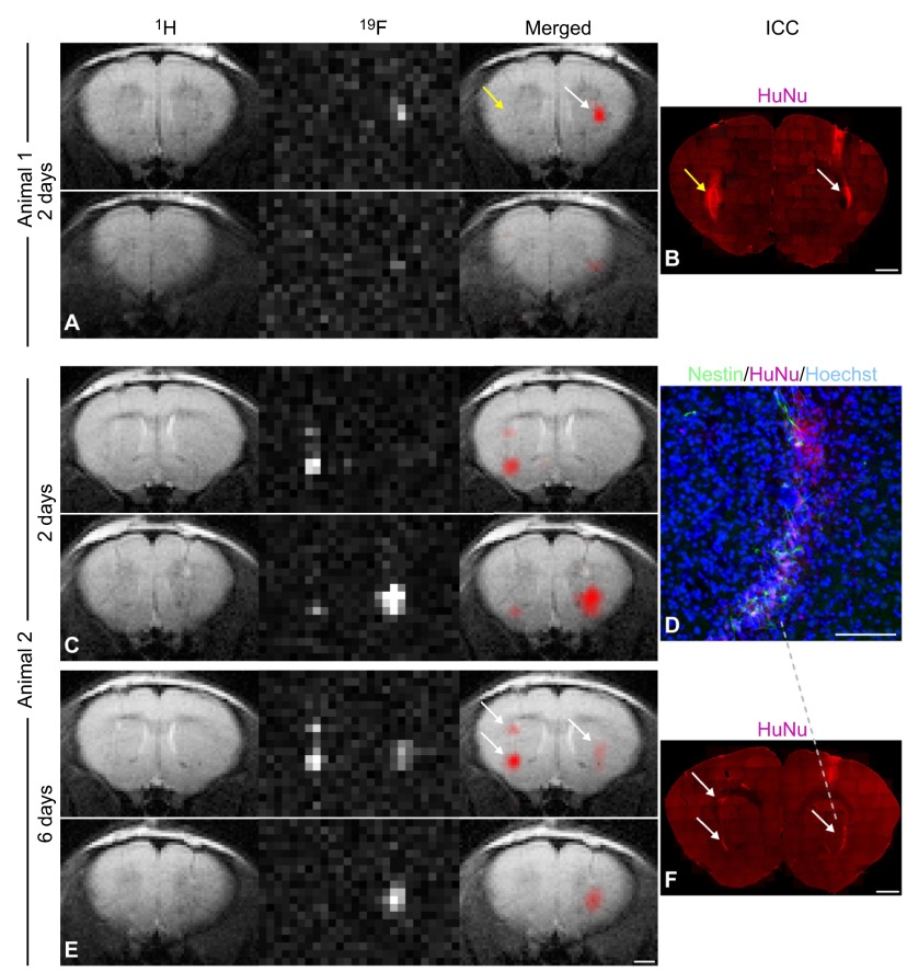
Fig. 2.
MRI of 19F-labeled cells in mice. MRI tracking of human neural stem cells (NSCs) labeled with fluorine (19F) after implantation in the mouse striatum. Animal 1: (A) 1H, 19F and merged MR images of a mouse brain acquired 2 days after injection with non-labeled control cells into the left striatum (yellow arrow) and with 19F-labeled NSCs into the right hemisphere (white arrow). Shown are two sequential slices demonstrating that only the labeled cells generated a 19F signal. (B) Human nuclear antigen (HuNu) staining was used to confirm the presence of NSC grafts in both hemispheres (yellow and white arrows). Animal 2: comparison of MR images of another mouse brain acquired (C) 2 days and (E) 6 days after grafting with human NSCs, showing no major signal loss in the 19F images over time. Shown are two sequential slices. This animal received two deposits of 19F-labeled cells in the left striatum and one deposit in the right striatum (E, arrows); the high spatial resolution of 19F MRI allows the two cell clusters in the left hemisphere to be clearly distinguished, demonstrating the potential of this technique for the detection of small numbers of cells in vivo within a restricted area. (D) Implantation of NCSs was confirmed histologically via colocalization of HuNu, the stem-cell marker nestin and the nuclear marker Hoechst in the mouse striatum. (F) HuNu staining correlated with the location and intensity of 19F signal generated from cell clusters (arrows). Scale bars: 50 μm for D; 1 mm for all others. Figure and data reproduced with permission (Boehm-Sturm et al., 2011).

