Fig. 5.
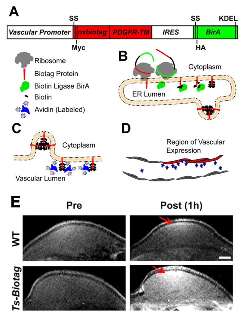
Schematic of the Biotag reporter system. The Biotag reporter system, as reported by Bartelle and colleagues (Bartelle et al., 2012), was used here to label endothelial cells in a mouse model of angiogenesis. This system is based on the coexpression and interaction of a Biotag and BirA, and in this study the authors relied on the strong affinity between avidin and biotin. (A) The authors generated transgenic mice that express multiple (nx) copies of a ‘Biotag’ (shown in red). The Biotag was fused to a signal sequence (SS; targeting to the secretory pathway) and the Myc-tagged transmembrane (TM) domain of the platelet-derived growth factor receptor (PDGFR-TM), followed by an internal ribosome entry site (IRES) sequence (allowing translation initiation in the middle of the mRNA sequence) to co-express the hemagglutinin (HA)-tagged BirA enzyme (which is a biotin ligase), modified with SS and KDEL (Lys-Asp-Glu-Leu amino acid) sequences. The expression of this bicistronic construct can be driven by different vascular endothelial cell promoters to restrict its expression to specific cell subtypes. (B) On translation, the ribosome recognizes the N-terminal SS of each protein to insert them into the lumen of the endoplasmic reticulum (ER). In the ER, the BirA enzyme (green) ligates free biotins (black rectangles) onto the Biotag protein (red) and, by continuing through the secretory pathway, the resulting biotinylated Biotag protein will be expressed on the surface of endothelial cells, whereas the BirA enzyme is retained in the ER by the KDEL sequence. (C) Thanks to the strong affinity between biotin and avidin, once on the cell surface, the biotins bound to the Biotag protein will be accessible for binding to the avidinated probes, which can be labeled with fluorescent or paramagnetic agents. (D) Based on the labeling, avidinated probes can be imaged via fluorescence or MR imaging, allowing for the selective detection of the vasculature endothelial cells (red) that express the transgene. (E) To image vascular endothelial cells in vivo, transgenic Ts-Biotag mice were generated by using a minimal promoter for tyrosine-protein kinase receptor 2 (T-short; Ts). After injection of DTPA-gadolinium-labeled avidinated probes, Ts-Biotag plugs could be visualized via MRI. Here, in vivo MR images of angiogenic endothelial cells within a Matrigel pellet doped with vascular endothelial growth factor, acquired before and 1 hour after injection, are shown (n=4). After injection, the immediate vascular tissue close to the plugs is clearly enhanced (arrow) compared with wild type (WT; n=4). Figure and data reproduced and modified with permission (Bartelle et al., 2012). Scale bar: 1 mm.

