Fig. 2.
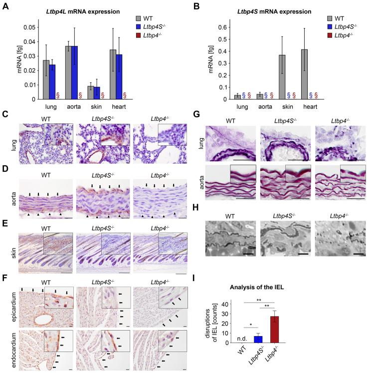
Localization patterns of Ltbp-4L, Ltbp-4S and elastin. (A) Quantitative PCRs of lung, aorta, skin and heart of WT and Ltbp4S−/− mice showing that each tissue had different amounts of Ltbp4L mRNA. There was no mRNA expression of Ltbp4L in lung, aorta, skin and heart of Ltbp4−/− mice (n≥3; §, no expression detectable). (B) Quantitative PCRs of lung, aorta, skin and heart of WT mice showing that each tissue had different amounts of Ltbp4S mRNA. There was no mRNA expression of Ltbp4S in lung, aorta, skin and heart of Ltbp4S−/− and Ltbp4−/− mice (n≥3; §, no expression detectable). (C) Representative images of Ltbp-4 immunoreactivity of lungs from WT, Ltbp4S−/− and Ltbp4−/− mice. Ltbp-4 was localized particularly in bronchial and bronchiolar walls and in vascular walls of WT and Ltbp4S−/− mice. Lungs of Ltbp4−/− mice were negative for Ltbp-4 immunoreactivity. Scale bars: 20 μm. (D) Representative images of Ltbp-4 immunoreactivity of aortas from WT, Ltbp4S−/− and Ltbp4−/− mice. Black arrows point to the aortic luminal side and black arrowheads to the adventitia. Ltbp-4 immunoreactivity was present in the vicinity of aortic elastic lamella throughout the entire aorta (from the endothelial lining to the adventitia) of WT mice and in the vicinity of the internal elastic lamella (IEL) and in the adventitia of Ltbp4S−/− mice. The aortic intramural elastic lamella of Ltbp4S−/− mice and the entire aorta of Ltbp4−/− mice showed no immunoreactivity for Ltbp-4. Scale bars: 20 μm. (E) Representative images of Ltbp-4 immunoreactivity of skin from WT, Ltbp4S−/− and Ltbp4−/− mice. In WT skin, Ltbp-4 immunoreactivity was present in the entire dermis, whereas it was completely absent in the epidermis. There was no difference in the tissue distribution of Ltbp-4 between WT and Ltbp4S−/− skin. The skin of Ltbp4−/− mice expressed no Ltbp-4. Scale bars: 20 μm. (F) Representative images of Ltbp-4 immunoreactivity of hearts from WT, Ltbp4S−/− and Ltbp4−/− mice. Upper panel: black arrows point to the epicardium of the heart. Ltbp-4 immunoreactivity was present within the myocardium and in the epicardium of WT mice. The myocardium and the epicardium of Ltbp4S−/− and Ltbp4−/− mice were negative for Ltbp-4 immunoreactivity. Lower panel: black arrows point to the endocardium. The endocardium of WT and Ltbp4S−/− mice clearly has Ltbp-4 immunoreactivity, whereas Ltbp4−/− mice were negative for Ltbp-4 immunoreactivity. Scale bars: 20 μm. (G) Representative histochemical elastica stainings of lungs (upper panels) and aortas (lower panels) displaying moderate elastic fiber fragmentation with intact and disrupted elastic fibers in Lbp4S−/− mice compared to WT mice and an increased degree of fragmentation of the elastic fibers in Ltbp4−/− mice compared to Lbp4S−/− mice. Scale bars: 20 μm. (H) Representative semi-thin sections of lungs showing elastic fibers with fragmented and intact parts in Lbp4S−/− mice and total disruption of elastic fibers in Ltbp4−/− mice compared to WT mice. Scale bars: 6 μm. (I) Quantitative analysis of disruptions of the IEL showing significantly higher numbers of disruptions in Ltbp4−/− mice compared to WT and Ltbp4S−/− mice and significantly higher numbers of disruptions in Ltbp4S−/− mice compared to WT mice (n=6; n.d., not detectable; *P<0.05, **P<0.01).

