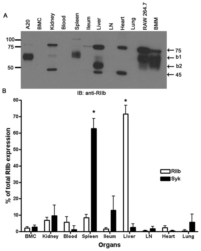
Figure 1. Most RIIb of mouse is in liver.
A. An ECL-developed immunoblot using rabbit anti-mouse RIIb antibody showing RIIb expression in several tissue and cell lysates prepared as described in M&M. Numbers are MW markers in kDa. B. Bar graph expressing the means and standard deviations of immunoblot-derived band densities, after factoring total organ weight for both RIIb isoforms and Syk from all organs (n=3 WT mice). The asterisks indicate that the expression levels of Syk and RIIb in spleen and liver, respectively, are statistically significantly different from the average of all other organs (P<0.001) (See M&M).

