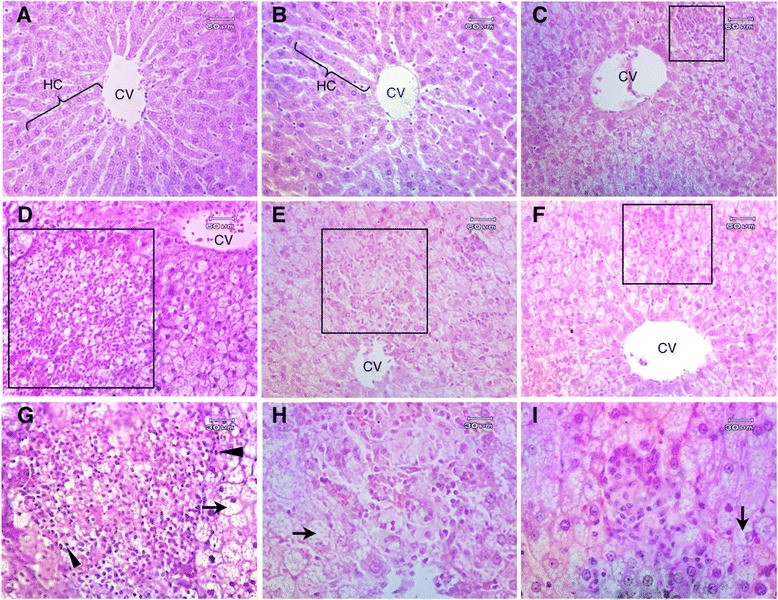
Figure 3.

Microscopic characters of liver tissue. Liver tissue of normal rats treated with distilled water (A) and 2.0 g/kg/day of RYR extract (B), and liver tissue from hypercholesterolemic rats treated with 5.0 mg/kg/day of rosuvastatin (C), distilled water (D, G), 1.0 g/kg/day of RYR extract (E, H), and 2.0 g/kg/day of RYR extract (F, I). Original magnifications were 100x (A-F) and 200x (G-I). Lipid deposition in hepatocyte (steatosis) (arrow), steatosis with inflammatory cell infiltrations (steatosis hepatitis) (quadrilateral) and nuclear condensation (head arrow) were observed in the liver of hypercholesterolemic rats (D-I). No remarkable damage was detected in SP and SP-2 g (A, B). CV, central vein; HC, hepatic cord (H & E).
