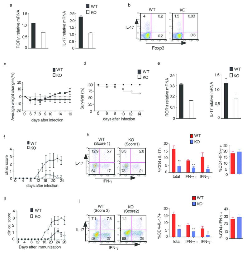
Fig. 6. E-proteins regulate Th17 responses in vivo.
(a,b) ER-cre-positive and WT mice were administered tamoxifen by gavage five times every other day. Four weeks after the final tamoxifen administration, lamina propria cells were purified from the small intestine; some of the cells obtained were directly analyzed by RT-PCR for RORγt or IL-17 (a), some of the cells were stimulated with PMA/Iono for 4hrs with Golgi stop in the last 3hrs and then analyzed for IL-17-expressing cells by flow cytometry gated on CD4+ cells (b). Data are representative of two independent experiments
(c,d) ER-cre-positive and WT were were orally administered with tamoxifen every other day for five times, One week after the final dose, mice were orally inoculated with C. rodentium and were weighed at the indicated time points (d) and followed for survival. Data are representative of two independent experiments.
(e) Colonic tissue obtained from WT or KO infected mice at 14 days after initiation of infection were subjected to real-time RT-PCR analysis of RORγt and IL-17 expression. Data are representative of two independent experiments.
(f) EAE disease course in WT and E2Af/+HEBf/f Cre+(KO) mice that had been treated with tamoxifen as above. Data are representative of two independent experiments. n=5(WT), n=6(KO).
(g) EAE disease course in Rag2-deficient mice reconstituted with 20×106 purified CD4+ T cells from WT and E2Af/+HEBf/f Cre+(KO) mice that had been treated with tamoxifen as above. n=6(WT), n=6(KO). Data are representative of two independent experiments.
(h) Cytokine production by cells isolated from the spinal cords of WT and E2Af/+HEBf/f Cre+(KO) mice on day 23 after EAE induction. The cells were stimulated for 4hr with PMA/Iono in the presence of Golgi stop and then analyzed for surface and intracellular cytokines by flow cytometry gated on CD4+ cells. Clinical score are shown in parentheses. Data are representative of two independent experiments. Tabulated Results from 2 independent experiments: Total: all IL-17+ cells; IFN-γ+: IL-17+IFN-γ+ cells; IFN-γ−:IL17+IFN-γ− cells. Error bars represent standard deviation; ** p<0.02, * p<0.05
(i) Cytokine production by cells isolated from the spinal cord of Rag2-deficient mice reconstituted with tamoxfen-treated WT or KO purified CD4+ T cells on day 20 after EAE induction. The cells were stimulated for 4hr with PMA/Iono in the presence of Golgi stop and then analyzed for surface and intracellular cytokines by flow cytometry gated on CD4+ cells. Clinical scores are shown in parentheses. Data shown are representative of two independent experiments. Tabulated results are from two independent experiments; Total: all IL-17+ cells; IFN-γ+: IL-17/IFN-γ+cells; IFN-γ−:IL17+IFN-γ− cells. Error bar represent standard deviation; ** p<0.01, *p<0.05

