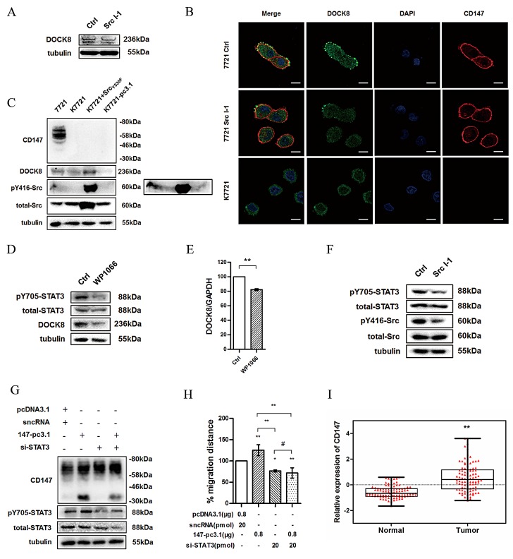
Fig.6. Src promotes DOCK8 expression via enhancing STAT3 phosphorylation.
(A) DOCK8 expression was detected using western blotting after treatment of 7721 cells with Src I-1. (B) Confocal microscopy images of 7721, 7721 treated with Src I-1 and K7721 cells. Red: CD147; Green: DOCK8; Blue: DAPI. Scale bar = 20μm. (C) DOCK8 expression and Src activity were examined in total lysates of 7721, K7721, K7721 transfected with SrcY530F and K7721-pcDNA3.1 cells (K7721 cells were transfected with pcDNA3.1 vector as a mock control for R7721) using western blotting. Phosphorylation of Src at Tyr416 can also be seen in the overexposed panel (right).(D) Phosphorylation level of STAT3 and DOCK8 expression were detected using western blotting after treatment of 7721 cells with WP1066. (E) The DOCK8 mRNA level was detected using real-time PCR after treatment of 7721 cells with WP1066. (F) Phosphorylation level of STAT3 was detected using western blotting after treatment of 7721 cells with Src I-1. (G) Phosphorylation level of STAT3 and CD147 expression were determined in 7721 cells overexpressing CD147 and/or transfected with STAT3 siRNA. (H) The effects of CD147 overexpression and/or STAT3 silencing on cell motility of 7721 cells. (I) CD147 expression using normalized microarray gene expression data. The bars represent each sample performed in triplicate, and the error bars indicate ± SD. * P < 0.05, ** P < 0.01, # P > 0.05, by unpaired t-test (E), one-way ANOVA (H) and paired t-test (I).

