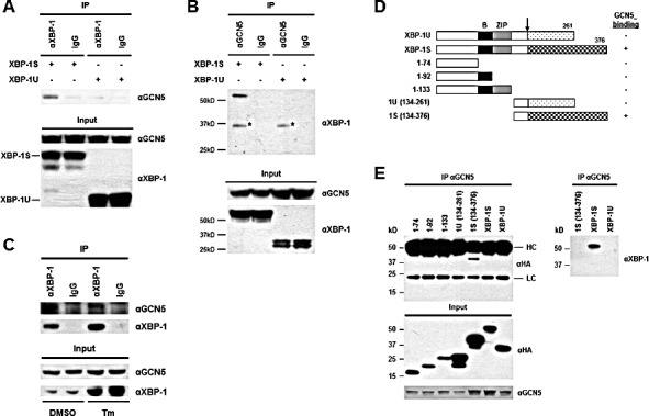
Figure 1. XBP-1S interacts with GCN5 through its specific C-terminal region.

(A) 293T cells were co-transfected with a GCN5 and an indicated XBP-1 expression plasmid (XBP-1U or XBP-1S). IP was performed by incubating the cell lysates prepared from the transfected cells with anti-XBP-1 (A) or anti-GCN5 (B) antibodies. Normal IgG (IgG) was used as a negative control and non-specific protein bands were marked with asterisks. The immunoprecipitated complexes and the protein inputs were analyzed by Western blotting. (C) Nuclear extracts prepared from cells treated with or without Tm were analyzed IP using an anti-XBP-1 antibody. Tm was dissolved in DMSO and the final concentration of DMSO in the culture was kept at 0.1%. Cells treated with 0.1% DMSO were served as a negative control. The immunoprecipitated complexes and the protein inputs were analyzed by Western blotting. (D) Diagram of XBP-1 truncations. All the constructs were HA-tagged. B, basic domain; ZIP, leucine zipper domain. (E) 293T cells were co-transfected with a GCN5 and an indicated plasmid to express an individual XBP-1 deletion. IP was performed using the anti-GCN5 antibody followed by Western blotting with anti-HA or anti-XBP-1 antibodies.
